Technology

Modelling Technology Background
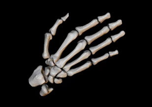
3-dimensional statistical models of shape and appearance are at the heart of image analysis at Imorphics. Our team of imaging scientists has been applying statistical models to medical image analysis for over 20 years, after originally publishing methodology representing shape as a statistical model at the University of Manchester in 1993.
Biological objects such as bones, brains and kidneys have characteristic shapes and appearance, but also show variation from person to person. Statistical models capture both the common features and the variation of a tissue by training the model with a representative set of images. During training key information is automatically extracted and stored, along with information on how to find the tissue in images from people not in the training set. Once this is done, the model can be used to automatically identify the tissue in similar images, look for changes in tissue during drug trials, or to examine differences between tissues with and without a particular disease.